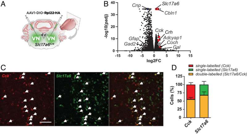
Identification of glutamatergic neuronal subpopulations in the VN. (A) Slc17a6Cre mice were bilaterally injected in the VN with a viral vector expressing the RiboTag (AAV1-DIO-Rpl22HA) for the molecular profiling of VGLUT2VN neurons. (B) Differential expression analysis showing significant enrichment for candidate VGLUT2VN neuron subpopulation marker transcripts in the IPs of RiboTag assays when compared to inputs (n=3). Specific enrichment for Slc17a6 (Vglut2), and depletion for inhibitory neuron (Gad2) and non-neuronal marker transcripts (Cnp, Gfap) were confirmed in the analysis (padj: adjusted P-value; FC: fold change). (C) Double-label in situ hybridization assay showing expression of Cck mRNA within Slc17a6-expressing cells. (Scale bar: 100 µm.) (D) Percentage of double-labeled Cck– and Slc17a6-expressing cells. Credit: Proceedings of the National Academy of Sciences (2023). DOI: 10.1073/pnas.2304933120
A team of neuroscientists at Universitat Autònoma de Barcelona has found that the neurons in a mouse brain’s vestibular nuclei control the signals that cause motion sickness. In their paper published in Proceedings of the National Academy of Sciences, the group describes how they exposed mice to bursts of spinning to give them motion sickness and then tested protein production in certain parts of their brains.
Prior research has suggested that motion sickness in humans is due to a disconnect between the way the brain processes visual data and inner ear signaling—the results are often nausea and vertigo. In this new effort, the researchers investigated what goes on in the brain when a mouse is experiencing motion sickness.
They attached a tube to the top of a spinning device and then placed test mice into the tube. The mice were spun for short bursts after which they were offered a meal. If a mouse refused the meal, it was deemed to be experiencing motion sickness and was ready for further testing.
Testing of the mice consisted of measuring levels of the protein VGLUT2 produced in their brains—it is produced by vestibular nuclei, which are neurons in the inner ear. Prior research has shown that such neurons are used by the brain to help maintain balance. The researchers found heightened levels of VGLUT2 in the mice after being subjected to the spinning device. This they reasoned, led to the symptoms evident in motion sickness.
To test their theory, they injected test mice that had not been spun with VGLUT2 and found that they exhibited the same symptoms as those that had been spun, including loss of appetite. Taking a closer look at the protein, the team found that a subset of VGLUT-expressing vestibular nuclei neurons that incited expression of the genes responsible for encoding a hormone called cholecystokinin (CCK) were responsible for most of the motion sickness symptoms.
The team then injected spun mice with a drug that blocked production of the CCK-A receptor and found that doing so resulted in greatly reduced symptoms. They conclude by suggesting their findings could result in the development of new therapies to treat motion sickness in humans and in the development of a new kind of appetite suppressant.
More information:
Pablo Machuca-Márquez et al, Vestibular CCK signaling drives motion sickness–like behavior in mice, Proceedings of the National Academy of Sciences (2023). DOI: 10.1073/pnas.2304933120
© 2023 Science X Network
Citation:
Experiments on mice reveal the parts of the brain involved in motion sickness (2023, October 19)
retrieved 19 October 2023
from https://medicalxpress.com/news/2023-10-mice-reveal-brain-involved-motion.html
This document is subject to copyright. Apart from any fair dealing for the purpose of private study or research, no
part may be reproduced without the written permission. The content is provided for information purposes only.
>>> Read full article>>>
Copyright for syndicated content belongs to the linked Source : Medical Xpress – https://medicalxpress.com/news/2023-10-mice-reveal-brain-involved-motion.html


