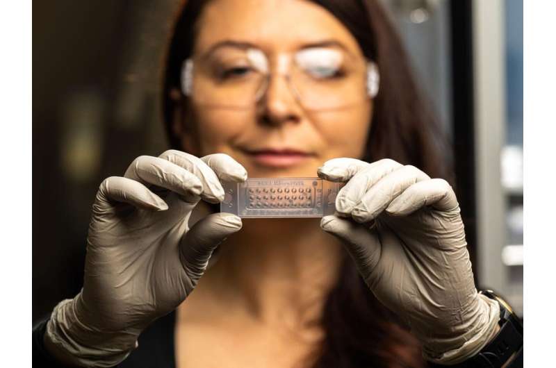
MicroPOTS is a chip-based approach developed by Pacific Northwest National Laboratory that allows sample processing in a microliter droplet to greatly improve the sensitivity of proteomics measurements. Credit: Andrea Starr | Pacific Northwest National Laboratory
Human tissue is composed of distinct cell types arranged in complex structures that send protein signals both within a single cell and between cells. In standard proteomics measurements, these cell structures mix to blend this network of signals together.
Scientists at Pacific Northwest National Laboratory (PNNL) used stained human pancreas samples to enable visualization of the islet (insulin producing micro-organs) regions of the pancreas and showcase how the microPOTS proteomics technology can be used to characterize region-specific protein signals.
This approach revealed insulin signaling proteins within the human pancreas samples were localized around the islet cells, not farther away. Their study is published in Molecular & Cellular Proteomics.
Currently, most ‘omics measurements derived from genes, transcripts, proteins, or metabolites come from bulk tissue. However, when scientists measure an entire tissue segment, microscopic changes that occur across the biological specimen are often missed.
The study demonstrates how standard analysis pipelines can be used on spatial proteomics measurements, enabling researchers to identify trends across the tissue that cannot be measured at the bulk tissue level. By applying these computational tools to the microPOTS data, biological signaling pathways that are altered within a specific area of the tissue can be identified. This finding proves that these computational approaches can be used to interpret additional spatial proteomics measurements and identify novel biology.
The need for a clinically accessible method to match protein activity within heterogeneous tissues is currently unmet by existing technologies. MicroPOTS can be used to measure relative protein abundance in micron-scale samples alongside the spatial location of each measurement, thereby tying biologically interesting proteins and pathways to distinct regions. However, given the smaller pixel/voxel number and amount of tissue measured, standard mass spectrometric analysis pipelines have proven inadequate.
The research adapts existing computational approaches to answer specific biological questions asked in spatial proteomics experiments. This new approach presents an unbiased characterization of the human islet microenvironment comprising the entire complex array of cell types involved while maintaining spatial information and the degree of the islet’s sphere of influence.
Researchers identify specific functional activity unique to the pancreatic islet cells and demonstrate how far their signature can be detected in the adjacent tissue. Results show that pancreatic islet cells can be distinguished from the neighboring exocrine tissue environment, recapitulate known biological functions of islet cells, and identify a spatial gradient in the expression of RNA processing proteins within the islet microenvironment.
More information:
Sara J.C. Gosline et al, Proteome Mapping of the Human Pancreatic Islet Microenvironment Reveals Endocrine–Exocrine Signaling Sphere of Influence, Molecular & Cellular Proteomics (2023). DOI: 10.1016/j.mcpro.2023.100592
Citation:
Chemical and computational advances enable 2D proteomics measurements of spatial signals (2024, February 5)
retrieved 5 February 2024
from https://medicalxpress.com/news/2024-02-chemical-advances-enable-2d-proteomics.html
This document is subject to copyright. Apart from any fair dealing for the purpose of private study or research, no
part may be reproduced without the written permission. The content is provided for information purposes only.
>>> Read full article>>>
Copyright for syndicated content belongs to the linked Source : Medical Xpress – https://medicalxpress.com/news/2024-02-chemical-advances-enable-2d-proteomics.html


