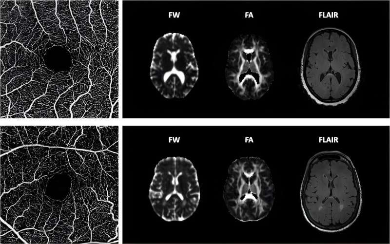
Optical coherence tomography angiography (OCTA) images representative of high (top row), and low (bottom row) OCTA perfusion measures, and their corresponding FW (free water), FA (fractional anisotropy), and FLAIR (fluid attenuated inversion recovery) images from brain magnetic resonance imaging scans. Credit: Alzheimer’s & Dementia (2023) DOI: 10.1002/alz.13469.
A research team led by the Keck School of Medicine of USC has discovered that a non-invasive eye exam may be a possible tool for screening Black Americans and other people from underdiagnosed and high-risk populations for cerebral small vessel disease, a major contributor to cognitive impairment and dementia. After Alzheimer’s disease, vascular dementia, associated with impaired blood flow to the brain, is the second most common dementia diagnosis.
“Most people with cerebral small vessel disease are not diagnosed until significant brain damage has occurred. Damage to the brain cells is not reversible,” said Xuejuan Jiang, Ph.D., associate professor of ophthalmology at the Keck School of Medicine and lead author of the study that was published in the journal Alzheimer’s & Dementia “This exam might help identify those people who are at high risk of developing cerebral small vessel disease early, while they can still get help.”
Using a new type of device that looks at the blood vessels in the retina, the team was able to connect certain characteristics in the vasculature of the eye to early signs of cognitive decline and structural changes commonly found in the brains of people with cerebral small vessel disease.
New imaging, new insights
Recruited from the African American Eye Disease Study, a population-based study of more than 6,000 African Americans from Inglewood, California, the research participants were all over 40 and had no history of cognitive impairment. Study participants underwent a type of retinal imaging called optical coherence tomography angiography, or OCTA, which was carried out at the USC Roski Eye Institute.
OCTA, a relatively new type of imaging that is increasingly incorporated in clinical practice in ophthalmology, captures detailed images of tiny retinal capillaries without needing to inject a dye into the patient. Using these images, the team was able to calculate the density of these blood vessels within the retina, the amount of blood that is flowing through those vessels and how quickly the blood was moving. Jiang noted that OCTA can capture changes in retinal capillaries before patients have clinical symptoms.
Participants were put through a series of examinations to evaluate their cognitive function. Some of them also underwent magnetic resonance imaging (MRI) of the brain which allowed the team to take measurements of the participants’ brain structures. Certain early structural changes in the brain are known biomarkers of cerebral small vessel disease.
A common connection
After analyzing the information collected during the exams, the team found that lower rates of blood flow through the retinal capillaries and lower density of blood vessels are both connected to functional and structural changes in the brain associated with cerebral small vessel disease.
Among people in the study who underwent cognitive testing, lower rates of blood flow and blood vessel density were connected to worse information processing speed and executive function. They were also associated with three MRI measures which are known to be related to cerebral small vessel disease.
“This indicates that altered retinal blood flow may be a biomarker of early changes in cognition resulting from cerebral small vessel disease,” said Jiang, noting that of the two associated measures, the rate of blood flow in the retinal capillaries was a more sensitive measure of changes in the brain. Jiang noted that this technology might also be used to monitor the progression of disease or the efficacy of treatments for cerebral small vessel disease.
Focus on diversity
It is also notable, said Jiang, that the study was carried out with Black participants because Black people typically have not been included in significant numbers in the research and clinical trials related to dementia and Alzheimer’s disease, despite dementia being more prevalent among Black people than white people in the United States.
It is critical that studies on cerebral small vessel disease and vascular dementia, noted Jiang, include diverse participants because these conditions are more commonly found among populations of people with high rates of diabetes, hypertension and other vascular diseases, which includes Blacks and Latinos.
“We know how important it is for research to include more diverse patients,” said Jiang. “There needs to be more research on Blacks and Latinos because they are at higher risk, and we are hopeful that this research is moving in the direction of finding a screening and monitoring tool.”
More information:
Retinal perfusion is linked to cognition and brain MRI biomarkers in Black Americans, Alzheimer’s & Dementia (2023) DOI: 10.1002/alz.13469. alz-journals.onlinelibrary.wil … ll/10.1002/alz.13469
Citation:
Study discovers possible tool to diagnose common contributor to vascular dementia (2023, October 6)
retrieved 6 October 2023
from https://medicalxpress.com/news/2023-10-tool-common-contributor-vascular-dementia.html
This document is subject to copyright. Apart from any fair dealing for the purpose of private study or research, no
part may be reproduced without the written permission. The content is provided for information purposes only.
>>> Read full article>>>
Copyright for syndicated content belongs to the linked Source : Medical Xpress – https://medicalxpress.com/news/2023-10-tool-common-contributor-vascular-dementia.html
