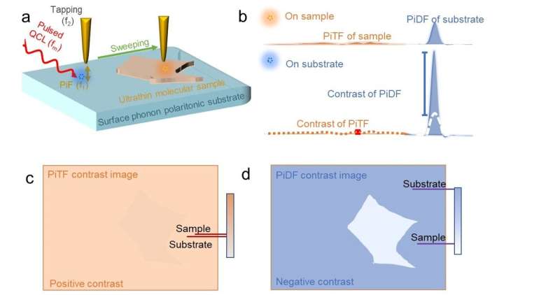
Phonon polariton enhanced nano-IR contrast imaging platform. (a) Sketch of the substrate-enhanced nano-IR contrast imaging platform based on a polar crystal substrate under the metallic tip. (b) Typical PiFM spectra were observed on the sample near its IR resonance and on the substrate near the tip-induced nearfield resonance. (c, d) Schematics for imaging of (c) PiTF of sample and (d) PiDF of the substrate and on a layered sample deposited on a PiDF-dominant substrate as depicted in (a). Credit: National Science Review (2024). DOI: 10.1093/nsr/nwae101
A new study has been led by Prof. Xing-Hua Xia (State Key Lab of Analytical Chemistry for Life Science, School of Chemistry and Chemical Engineering, Nanjing University). While analyzing the infrared photoinduced force response of quartz, Dr. Jian Li observed a unique spectral response that is different from the far field infrared absorption spectrum.
“The photoinduced force response follows the real part rather than the imaginary part of the dielectric function of quartz.” Dr. Li says. “We immediately discussed with the theorist Dr. Junghoon Jahng to analyze the experimental results, and we agreed that it is the quartz’s unique surface phonon polariton that extremely enhances the photoinduced dipole force.”
A paper describing these findings is published in the journal National Science Review.
To verify this result, the team compared the spectral response of quartz using photothermal induced resonance (PTIR) and photoinduced force microscopy (PiFM), showing that photoinduced dipole force (PiDF) dominates the photoinduced thermal forces (PiTF) of quartz. As PiDF shows a more pronounced relationship with the tip-quartz distance (~z−4) compared to the PiTF ( ~z−3), Dr. Li proposed a general approach for nano-IR contrast imaging of ultrathin samples loaded on top of quartz.
The ultrathin sample, characterized by a positive real part of the permittivity (weak oscillator), is expected to manifest weak PiTF and PiDF near its infrared (IR) resonance. However, a significant PiDF change is anticipated near the tip-induced nearfield resonance of the quartz substrate.
These spectral distinctions contribute to the contrasts in nano-IR imaging. Notably, the PiDF response on quartz exhibits a more conspicuous signal variation with respect to sample thickness compared to the PiTF of the sample. For ultrathin samples, PiDF imaging on quartz presents an opposite contrast with enhanced sensitivity compared to the nano-IR contrast imaging with the PiTF of the sample.
The team used a polydimethylsiloxane (PDMS) wedge prepared on a quartz substrate to demonstrate the substrate-enhanced nano-IR contrast imaging. The results provide clear evidence that the PiDF can be employed for sensitive nano-IR imaging of ultrathin samples under nanocavity geometry with improved contrast and sensitivity.
The researchers further applied the nano-IR imaging method to visualize thin covalent organic framework layers and subsurface defects under block-copolymer films. They hypothesized that by selecting suitable IR materials that exhibit phonon polaritons/reststrahlen bands, users could achieve high-resolution nanoimaging of specific crystals and polymer molecules, as well as biomolecules with known vibrational mode frequencies.
More information:
Jian Li et al, Surface-phonon-polariton-enhanced photoinduced dipole force for nanoscale infrared imaging, National Science Review (2024). DOI: 10.1093/nsr/nwae101
Citation:
Ultrathin samples with surface phonon polariton enhance photoinduced dipole force (2024, May 6)
retrieved 6 May 2024
from https://phys.org/news/2024-05-ultrathin-samples-surface-phonon-polariton.html
This document is subject to copyright. Apart from any fair dealing for the purpose of private study or research, no
part may be reproduced without the written permission. The content is provided for information purposes only.
>>> Read full article>>>
Copyright for syndicated content belongs to the linked Source : Phys.org – https://phys.org/news/2024-05-ultrathin-samples-surface-phonon-polariton.html


